The National Eye Institute (NEI) researchers have conducted a study for preventing vision loss from human diseases such as retinitis pigmentosa. According to a new study silencing a gene called Nrl in mice prevents the loss of cells from degenerative diseases of the retina.
The tissue in the back of the eye called the retina contains two types of cells that convert light into electrical signals sent to the brain. Rod photoreceptors enable vision in dim light, while cone photoreceptors enable color vision and the ability to see in well-lit conditions. Genetic mutations affect mostly rods, leading to night blindness such as that seen with retinitis pigmentosa. However, because rods also provide vital structural and nutritional support to cones, rod dysfunction or death can lead to cone degeneration and blindness.
In a series of studies, Anand Swaroop, Ph.D., chief of NEI’s Neurodegeneration and Repair Laboratory, and his team asked if saving rods could prevent the loss of cones, thus preserving daylight and color vision.
During development, Nrl determines whether a photoreceptor precursor cell becomes a rod or a cone. In previous studies, Swaroop and colleagues showed that mice engineered to lack the Nrl gene develop cone-only retinas. Other researchers showed that if Nrl expression is knocked out in rods of mature mice, the cells gain cone-like features and survive despite rod gene mutations.
“The evidence suggested to us that coaxing rods into becoming more cone-like by knocking out Nrl was a potential strategy for overriding mutations that would otherwise lead to rod degeneration,” said Swaroop. “Consequently, the neighboring cones would remain functional and viable.”
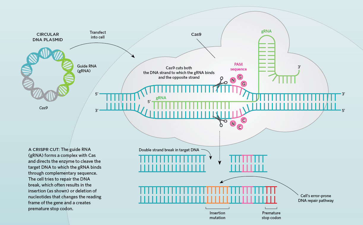
To test the strategy, the researchers used a new genome editing technology called CRISPR, which stands for clustered regularly interspaced short palindromic repeats. CRISPR is like a molecular pair of scissors that can be programmed to snip a specific DNA sequence with precision.
NEI postdoctoral research fellow and the study’s first author, Wenhan Yu, Ph.D., developed a method for applying CRISPR in photoreceptors. His strategy uses an adeno-associated virus (AAV) as a carrier, or vector, to introduce CRISPR into retinal cells. Yu tested this genome editing tool to remove the Nrl gene in wild-type mice and three different mouse models of retinal degeneration. By measuring gene expression and examining the retinal cells, the researchers confirmed that rods became more cone-like, as predicted. Although these cone-like rods could not detect light, they survived and improved survival of their neighboring cones.
In all three mouse models, rod degeneration was prevented or slowed, although less benefit was achieved when the therapy was introduced in older animals. Importantly, the benefit was evident in all three models, regardless of the specific gene defect in the mouse.
“Unlike conventional gene therapy, in which a normal gene is introduced to replace the defective gene, this approach could treat retinal degeneration caused by a variety of mutant genes,” explained Zhijian Wu, Ph.D., head of the NEI Ocular Gene Therapy Core and senior author of the study.
More research is needed before the therapy is ready for testing in a clinical trial. The safety of CRISPR has yet to be established and information is needed about its possible adverse effects. Nevertheless, these findings provide proof of concept for CRISPR-based therapies for degenerative retinal diseases.
The study was supported by the Intramural Research Program at NEI. NEI leads the federal government’s research on the visual system and eye diseases. NEI supports basic and clinical science programmes to develop sight-saving treatments and address special needs of people with vision loss.
Nih-funded scientists, deploy crispr, preserve photoreceptors in mice


22.jpg)

.jpg)